Understanding the role of vitamin K-dependent proteins in vascular calcification

What if the process of arterial calcification was regulated from within the cells of the blood vessels, and that it had nothing directly to do with what you ate and what circulated in the bloodstream because calcification takes place not anywhere near the surface but inside the blood vessel wall?

What if the process of arterial calcification was actually a process by which muscle is transformed into bone, a process by which vascular smooth muscle cells transform themselves into bone cells which then actually build bone tissue within the blood vessel wall?

And what if apoptosis preceded calcification, what if cell death was what triggered the process of calcification, and it was the apoptotic bodies of dead vascular smooth muscle cells within the blood vessel wall that served as the nodes around which calcium crystals formed?

Would you not find this shocking? Find it incredible that any of these could be true, let alone all of them? It’s entirely not at all what we’ve been told by “health experts” and “health authorities” for more than half a century!

All of these statements are hard to believe. It is especially unbelievable that muscle cells can change into bone-building cells, and begin to grow bone tissue within the artery wall. It sounds surreal, kind of like science fiction. But it isn’t. All of it is true. All of this has been observed.

Interesting, you may think, but what does any of this have to do vitamin K? Everything! It has everything to do with vitamin K.

How clever we are

The sophistication and precision of biochemical reactions and processes in animals and humans are mind blowing. Understanding how they work is a wonderfully noble endeavour that is certainly very fulfilling in its own right. In some cases though, it can be a matter of life and death. And in the case of the processes related to and regulated by vitamin K dependent proteins it definitely is.

This is not an exaggeration to push you to read on. It’s a statement of fact. And you’ll see how this is true by the time we finish. I believe it is essential, for each one of us to understand the details of how things in our body work and how they are related and connected in order to appreciate their significance and their importance.

We are so clever. We can figure out such complicated things when we put our minds to it. Things like complex biochemical pathways, or long chains of enzymatic reactions that, one step at a time, transform molecules from one form into another. And it is this kind of cleverness that has enabled us to develop the hundreds of different types of medications we can find today in drug stores.

We have designed medications to address basically every symptom we can think of. If it’s a symptom we’ve had, it’s most likely a symptom that many others have or have had. And if many have the same or a similar symptom, we can be sure that at least one pharmaceutical company will have made a drug for it.

Warfarin was developed in the 1950s to prevent or at least suppress coagulation, and in so doing help prevent or at least reduce the number of strokes and heart attacks. Because so many people either suffer from, are susceptible to, or are at risk of cardiovascular disease, many people take warfarin.

And what I mean by many in this case is between 20 and 30 million prescriptions per year in the United States alone. The number went up to 35 million in 2010 and dropped back to 20 million in 2015. That’s a lot of warfarin pills! You can see the stats here (http://clincalc.com/DrugStats/Drugs/Warfarin). Warfarin is in the top 50 drugs. It’s 42nd down the list. Just below aspirin at 39, insulin at 36, and ibuprofen at 34, as you can see here (http://clincalc.com/DrugStats/Top200Drugs.aspx).

Surely close to every household in the western world will have somewhere in a bathroom cupboard or drawer a bottle of aspirin or ibuprofen. Given how close to warfarin they are in popularity of usage, there’s clearly no need to even say that this anti-coagulant drug is in broad and widespread use.

Isn’t this great, though? Millions of people at risk of having blood clots that would possibly cause them a stroke or heart attack, protected by taking a little warfarin every day? Yes, I suppose in some ways, it is, if these people are actually at risk. But, unfortunately, with a drug like that, we can be pretty sure that most are taking it preventatively, as in, just in case. And this is a problem.

Warfarin works by disrupting the process that leads to the activation of coagulation factors. The blood’s ability to form clots quickly is one of its most vital functions, because without it we would just bleed to death from a flesh wound. Evolutionarily, we simply would not have made it to here without this protection mechanism that ensured that when we were wounded, the blood would immediately thicken to stop it bleeding out of our body by forming clots at the surface of the open wound as fast as possible. The special proteins responsible for regulating coagulation are vitamin K-dependent proteins (VKDPs).

It has taken a long time to understand, first of all, that there wasn’t just vitamin K, but in fact two different kinds of vitamin K. It is also true that it has taken a long time to identify the major vitamin K-dependent proteins and figure out how they work. We are talking about 40 years from the 1950s to the 1990s. So, you really shouldn’t be surprised if you haven’t read or heard about this before.

But today, a lot has been understood through in vitro and in vivo observations, trials and studies both in animal models and in humans. And even though we will inevitably continue to deepen our understanding of the subtleties of the molecular mechanisms, the species, and the interactions involved in the life of cells and proteins in how they affect the state of our blood vessels and organs, this is a sketch of the picture we have at this stage.

Vitamin K dependent proteins

There are about twenty identified VKDPs belonging to two classes: hepatic—those produced by the liver, and extra-hepatic—those produced in other tissues. Those from the first class are the most well-known and well-studied. They are the coagulation factors (II, VII, IX, and X) manufactured by the liver and activated within it before being pushed into the bloodstream and circulated throughout the body to maintain a healthy coagulation response in case it is ever needed. These are the ones targeted by warfarin. Naturally, since that drug has been around since the 1950s, the role and function of these vitamin-K dependent coagulation factors have also been known at least since that time.

The second class is less known and less studied but has—luckily for us—gained much more attention in the last two decades. It includes three very important proteins whose functions are essential in maintaining healthy blood vessels. But unlike the coagulation factors produced in the liver, these proteins are instead produced by the vascular smooth muscle cells and activated there locally in the vasculature. These vascular health factors, we call them that in analogy to but to distinguish them from the coagulation factors, were identified much more recently in the 1980s and 1990s. All are proteins that contain gamma-carboxyglutamic acid abbreviated Gla.

Some important ones for us here are osteocalcin, for which it took 30 years to be identified as an inhibitor of calcification when it was discovered in vitro to prevent the precipitation of crystals in a supersaturated calcium solution. This means that without it, calcium crystals would have inevitably formed spontaneously in the solution. Osteocalcin is also called bone Gla protein. Growth arrest specific protein 6 is involved in the regulation of cell proliferation, and seems to inhibit premature cell death. And the most important one in relation to soft tissue calcification, matrix Gla protein abbreviated MGP.

Matrix Gla protein was originally isolated from bone, but it has been found to be expressed in several other tissues including kidney, lung, heart, and—most critically—vascular smooth muscle cells or VSMCs. It is now known to be the most potent inhibitor of calcification of blood vessels, and even though the liver does produce and secrete MGP into the bloodstream, only the MGP produced in the vasculature inhibits calcification.

Besides being produced in different tissues, another important difference between the two classes of VKDPs is that the liver-produced coagulation factors are phylloquinone—or vitamin K1-dependent, whereas the vascular smooth muscle cell-produced proteins are menaquinone—or vitamin K2-dependent. In light of the fact that it is rather hard to find vitamin K1 insufficiency with a diet that contains at least some green plant foods, while the exact opposite is true for vitamin K2 of which the western diet is practically devoid, this difference is highly significant.

Both vitamin K1 and K2 are absorbed in the second and third portions of the small intestine, the jejunum and ileum, K1 is delivered to the liver, whereas K2 is transported via LDL and HDL to other organs. K1 is mainly found in the liver, whereas K2 is preferentially stored in peripheral tissues, with the highest levels in the brain, aorta, pancreas, and fat tissues. This obviously attests to the importance of these essential vitamins.

While vitamin K1 and K2 are really two different vitamins with different functions, transport mechanisms, and distribution in the tissues, and while there are several differences between the vitamin K1-dependent and the vitamin K2-dependent proteins, these have one essential thing in common. This is, as their name says, that they are vitamin K-dependent. What this means is that all these proteins share the same enzymatic chain of activation—whether it mediated by K1 or K2—that transforms them into their biologically active form, the form they need to have in order to do the things they are meant to do.

All VKDPs must be carboxylated in order to be activated. The process is complicated and not yet completely understood. We know that it is targeted to the glutamic acid (Glu) residues on the protein that must be made into gamma-carboxylglutamic acid (Gla). We also know that the process is mediated by the enzyme gamma-glutanyl carboxylase (GGC), and that vitamin K is the main co-factor that enables the enzyme to perform the activation. In the end, the process leads to the addition of a carbon dioxide molecule to the gamma-carbon of Glu, which transforms it into Gla. However, it is the reduced form of vitamin K that is required.

Vitamin K, whether it is the plant-based phylloquinone (K1) or the animal-based menaquinone (K2), enters the body through the diet in its non-reduced form. Reduction involves the addition of hydrogen in molecular form, H2, to make KH2. Transformations of this kind are generally always done by enzymes, and so is this one. In this case the enzyme is vitamin K epoxide reductase (VKOR). Its action is essential because it is the reduced form KH2 that acts as the co-factor in the process of carboxylation.

The energy released by the oxidation of KH2 drives the addition of the carboxyl group unto the glutamic acid residues. But the oxidised form of vitamin K, KO, can subsequently be reduced again to KH2. Thus vitamin K is first reduced, then oxidised to help push the carboxyl group unto the glutamic residue, and then reduced once more to start the whole cycle again. This cycle is called the vitamin K epoxide reductase or VKOR cycle.

For this class of proteins, the VKDPs, activation through carboxylation means for them to acquire the structure and properties needed to bind calcium in order to transport it. You may recall from a previous chapter in the story of vitamin K2, matrix Gla protein generally transports calcium out of soft tissues in order to prevent calcification, and bone Gla protein generally transports it into bones and teeth to prevent osteopenia, osteoporosis, and tooth decay.

The big red flag

Now you understand why it is that when, in our remarkable cleverness, we understood that the main coagulation factors depended on the action of these enzymes to be activated and rendered functional, we naturally concluded that the best way to prevent clot formation would be to prevent coagulation, and that this could be achieved by blocking these enzymes from doing what they are intended to in a healthy organism. This is precisely what warfarin does.

And it does it well. Otherwise it wouldn’t have become as commonly used as it is. And we can be certain it has saved a lot of people much of the pain and possibly life-threatening conditions that a blood clot could have caused them. The problem is that the vascular health factors so critical for maintaining healthy blood vessels, depend on the same enzymes for activation as do the coagulation factors. Preventing the carboxylation of coagulation factors, prevents, in exactly the same way, the carboxylation of the vascular health factors.

This was only understood to be a major problem relatively recently. We first had to understand that there isn’t just one kind of vitamin K, but that there are two, and that they are very different in their functions. We had to understand that both vitamin K1 and vitamin K2-dependent proteins rely on the same enzymes to get activated. We had to understand the carboxylation process by which they are activated. And we had to understand that MGP, BGP, and Gas 6 are vitamin K-dependent proteins, that they are specifically vitamin K2-dependent, how they are activated, what they actually do in our veins and arteries, and what happens if they can’t do what they are designed to do.

A major red flag about anticoagulants and warfarin came up from what was seen in mice. The first part of the study was with MGP-knockout mice, (mice in which the MGP-encoding gene was deactivated). They were observed to have stunted growth from the premature calcification of the epiphysis—the part at the end of bones and at the junction with the cartilage of the joint which allows the bone to grow longer. As as soon as the epiphysis calcifies, longitudinal growth stops. But this was the least severe of the problems that were observed.

The MGP-knockout mice very quickly developed severe arterial calcification, and died highly prematurely, within 6 to 8 weeks, of strokes, heart attacks, and rupture of the aorta. Normal lab mice live 2 to 3 years and some even up to 4 years. So, in the least extreme case, a MGP-knockout mouse dying from aortic rupture at 2 months instead of living a relatively short normal life of only 2 years, would be equivalent for a human that would normally live to the age of 72 to die at the age of 6!

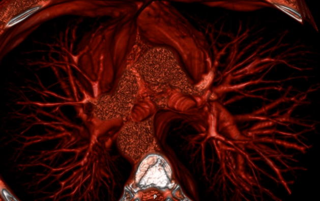
Here is what severe coronary calcification looks like in humans:

severe_coronary_calcification

Severe coronary calcification in a patient with end-stage renal disease. We can see that these blood vessels are basically filled with bone tissue that appears bright white. (https://www.bmj.com/content/362/bmj.k3887)

It was also observed that although the liver did produce and release MGP into the bloodstream, it had no effect on the arteries. Only the tissue-specific, locally-produced MGP within the vascular smooth muscle cells was able to inhibit calcification.

To check these conclusions, a similar study was done on normal mice that were given vitamin K1 to ensure proper liver function and healthy coagulation, and warfarin to block all extra-hepatic MGP action in tissues. The result? Stunted growth, pervasive arterial calcification, and premature death from stroke, heart attack, and aortic rupture.

The conclusions were solid: matrix Gla protein is the organism’s primary protection against soft tissue and arterial calcification; liver MGP has no protective effect on arteries, and only VSMCs-produced MGP can inhibit calcification in the arteries; both vitamin K deficiency and disruptions of the action of the enzymes that activate MGP cause extensive soft tissue calcification; and only vitamin K2, not vitamin K1, can inhibit warfarin-induced calcification.

Going further

When this was understood, more attention began to be paid to matrix Gla protein. Many other details were elucidated through further investigations. It was found that MGP is an 84-amino acid protein with five Gla residues. That all of these Gla residues are produced by gamma-carboxylation, which is mediated by the enzyme gamma-carboxylase that requires vitamin K2 as a cofactor, and that until now, the only known function of Gla residues is to bind calcium ions and crystals (calcium apatite). It was discovered that the concentration of calcium and phosphate in extracellular fluids is high enough to trigger and sustain growth of crystals, but that MGP and BGP prevent this from happening. That MGP is required by VSMCs to maintain their elastic and contractile nature. And not just that.

MGP actually inhibits the transformation of VSMCs into bone cells by antagonising the action of Bone Morphogenic Protein 2 (BMP2). It turns out that the muscle cells of the blood vessels have in them the potential to either stay smooth elastic contractile muscle cells, or turn into osteoblast-like bone building cells. BMP2 triggers that osteogenetic gene expression in the VSMCs: it tells muscle cells of the blood vessels to transform into bone-building cells.  And as if this wasn’t enough, BMP2 also induces apoptosis: it tells blood vessel muscle cells to commit suicide, which is certainly to help in the process given that once dead, they can be used as seeds for calcium crystal formation, and thus promote a faster and more efficient calcification.

What induces expression of BMP2 in cells? Probably several things that we haven’t yet identified. But for now we know that BMP2 is stimulated by oxidative stress, chronic inflammation, and high blood sugar levels. The good news is that MGP protects against all of these effects by antagonising BMP2. So if there is enough MGP and enough vitamin K2, if there are no disruptions to the action of the vitamin K dependent enzymes by anticoagulants like warfarin, and if oxidative stress, inflammation, and blood sugar are kept low, then there is protection against calcification of the arteries and other soft tissues like the liver, kidneys, and heart.

Recap

Here we have it. We have now understood the role of vitamin K dependent proteins in vascular calcification. And although it was a little long and maybe somewhat arduous, all the details are clear. It is complicated. I won’t deny that. But I have strived to make it all as accessible as I could without diluting the mechanisms of action and relationship between the different players. Let’s recap to make sure you are left with the essential elements in mind.

Vitamin K dependent proteins can either be vitamin K1 or vitamin K2 dependent. The dependence comes from the fact that vitamin K is required to activate the protein. This activation is the carboxylation in which a carbon dioxide is added to the glutamic acid residues along the protein. Carboxylation is mediated by carboxylase (GGC) that requires the reduced form of vitamin K in order to oxidise it and get the energy to push the carbon dioxide molecule onto the glutamic acid residue. Vitamin K is reduced by reductase (VKOR) which can do it over and over again in what is called the VKOR cycle.

Vitamin K1 dependent proteins are mostly liver based coagulation factors. Vitamin K2 dependent proteins are mostly outside the liver and generally involved in inhibiting soft tissue calcification. The most important calcification-inhibiting VKDP is matrix Gla (MGP), which performs a wide range of tasks to maintain elastic, flexible, calcium-free blood vessel walls.

Calcification is triggered by the death of vascular smooth muscle cells. These dead muscle cells act at seeds for calcium apatite crystals to form. VSMCs can be induced to become osteoblast bone-building like cells that then go on to stimulate the growth of bone tissue within the artery walls. This process is stimulated by bone morphogenic protein 2 (BMP2), which is expressed under conditions of oxidative stress, inflammation, and hyperglycaemia.

To prevent and reverse calcification the most important is to provide a good supply of vitamin K2 through diet and supplementation. Because it is essential in the activation of Gla proteins but only through its role in the VKOR cycle, the amount of K2 is the rate limiting factor. Hence more is better than less, and excess will simply remain unused but will not cause harm.

Naturally, matrix Gla protein needs to be available. Cells of tissues where calcification occurs (kidney, liver, heart, and blood vessels) secrete MGP. An interesting evolutionary self-protection adaptation mechanism. And here’s another: the amount of MGP that is produced by a cell depends on at least two factors that have been identified. One is the amount of calcium; the other is the amount of vitamin D3. In both cases, the more there is, the more MGP is produced.

So, vitamin D3 has the role of making calcium available but at the same time stimulates the production of MGP in order for the calcium to be available to the bones and not to the soft tissues. But for this, it relies on vitamin K2. This is why vitamin D3 without vitamin K2 leads to calcification: because MGP and BGP remain inactive and incapable of binding to the calcium ions to move them into bones and out of tissues. On the other hand, plenty of vitamin K2 would indeed activate the available MGP, but without enough vitamin D3 there might not be enough MGP to confer proper protection against calcification. This is a perfect example of the complementarity of action and function in essential micronutrients. There are certainly many more, but this one is particularly remarkable.

Final thoughts

I want to close on a final consideration. It is so easy and seems so natural for us to think in terms of this and that, good and bad, for and against, that our tendency is to look at everything in these terms. This is also true when we look at biochemical processes like the ones we have described and explored here. We naturally lean towards looking at the calcification inhibiting mechanisms as protective, and those that promote calcification and apoptosis as destructive.

But the reality is that cells, proteins, and enzymes don’t behave in these terms, they don’t think in these terms simply because they don’t think. They react biochemically to what they are exposed to, to the molecules and chemical messengers they encounter, to the quality of the liquids in which they bathe, to the characteristics of the environment in which they live, microsecond after microsecond, without any forethought or concern for the microsecond that will follow. The only guiding principle that can be used to lead us to understand why things happen the way they do is evolutionary adaptation to survive.

Having recognised this, we immediately see that the mechanisms that promote apoptosis of VSMCs, their subsequent transformation to osteoblast-like cells, and the growth of bone tissue within the artery walls that we refer to as arterial calcification, can only be a protection mechanism. A mechanism to protect the tissues and cells from the damaging effects of exposure to free radicals, inflammatory molecules, and glucose. Because, as we have seen, the process is reversible, it would be perfectly natural to undergo periods of calcification followed by periods during which the bone tissue is broken down and removed from our arteries and other soft tissues and organs when the circumstances allow it. Actually, we should say when the circumstances dictate it, because no matter what happens, it is always the circumstances—the environment—that dictate what is to happen.

What we can do, with the knowledge of what we have understood, is make choices about what we eat and drink, when and how much we eat, and how we live, sleep, and exercise. Choices that will shape or reshape, define or redefine the makeup of this internal environment of the body to always move us in the direction of optimal biochemistry, optimal physiology, optimal metabolism, and optimal health.

Everything that we explore together is always about just this. But sometimes the corrective action requires effort, sometimes even a lot of effort. In this case, however, it is as simple as can be, because it only requires us to supplement with vitamin K2 and possibly also D3. Of course, the last thing we want is a lifestyle that promotes the expression of BMP2 and the growth of bone tissue within our arteries. But supplementing with K2 and D3 together will in general bring only benefits. I know it was a very long-winded way to get to this, but now you understand why. That was—and is—the whole point of this blog, after all. I hope you enjoyed reading.


The information in this article comes primarily from the following papers: Molecular Mechanisms Mediating Vascular Calcification by Proudfoot and Shanahan (2006); Vitamin K-dependent Proteins, Warfarin, and Vascular Calcification by Danziger (2008); The Role of Vitamin K in Soft Tissue Calcification by Theuwissen, Smit, and Vermeer (2012).

Thank you to all our patrons, and in particular Eric Peters, for their continued support. Become a proud sponsor of healthfully and join our patrons today!

Rejecting the lipid hypothesis with a cholesterol of 278 mg/dl and a smile

When it comes to evaluating how likely you are to have a heart attack, the most accurate diagnostic—the gold standard—is the calcium score. The reason why it’s the most accurate is because it’s calculated from an actual 3D image of the heart and the blood vessels around it. A computerised tomography (CT) scan is done, and from it the amount of plaque buildup in all the places where it appears because of the high density of the calcium it contains is measured and summed to give the total calcium score.

3d_image_of_my_heart

3D volume rendering of my heart seen from the top.

Even though it has been estimated that approximately half of heart attacks are caused by non-calcified lesions, this is the closest thing we have to a direct measurement of the amount of plaque in the network of arteries around the heart. From doing this to thousands of people, we know that plaque usually begins to accumulate after the age of 35. Why isn’t the calcium score test done systematically on everyone above 40 in order to assess their immediate risk, but also to track their individual cardiovascular evolution, showing, with a reliable reference each year, how quickly or slowly arterial plaque is growing? Because it’s too expensive. Therefore, it’s only prescribed to people who are deemed to be at high risk based on other so-called “risk factors”. You know the list: overweight, sedentary, smoking, stressed, etc. But the clincher in this list of risk factors, the one factor that has pretty much eclipsed all the other ones, at least for the past few decades, is high cholesterol.

The focus on cholesterol was, over time, shifted to LDL, the “bad” cholesterol, and later on the ratio between it and HDL, the “good” cholesterol, terms introduced by the pharmaceutical industry to convince us that there is a battle between a good guy and a villain that must be stopped, which they can help with by providing us cholesterol lowering statins, even if with each passing year, the evidence exonerating cholesterol and lipoproteins from any wrong-doing in the genesis and progression of cardiovascular disease has been accumulating. Still, for people and for doctors, it’s really hard to overcome the several decades of conditioning we’ve suffered holding cholesterol as the main culprit for heart disease.

Fortunately, this knowledge and information have been shared and available for as long as the first experiments that set us on this damning direction in thinking and mindset. For my part, I first read a clear expose on the function of cholesterol and lipoproteins from Ron Rosedale over 10 years ago. Then I read it from Uffe Ravnskov, then from Anthony Colpo, then from Malcolm Kendrick who has and to this day continues to investigate the topic and share his findings on his blog, and then from Gary Taubes. All of this has taught me that cholesterol, HDL, and LDL, are not only not dangerous, but that they are essential and crucial for optimal health. This, I shared with you in But what about cholesterol? and shaped my diet to maintain healthy levels: I restricted carbohydrates and polyunsaturated oils, and have gotten most of my calories from minimally processed saturated fats from grass fed animals fats, coconut oil, butter, and olive oil. In this endeavour to maintain strong cholesterol and lipoprotein levels, as you can see below, I have succeeded.

The following plot shows all the measurements of total cholesterol I have ever gotten made from blood tests over the past decade. What you can see is that in late 2007—a time before which I ate mostly complex carbohydrates and polyunsaturated seed oils while avoiding animal and saturated fats—my total cholesterol was below 150 mg/dl. Since then, it has been generally around or above 200 mg/dl with a slight upward trend over the years.

ts_total

My own total cholesterol levels in mg/dl measured from late 2007 to mid 2018.

If we look at the concentration of low and high density lipoproteins LDL and HDL, we also see consistently high levels, with LDL typically 10-30 mg/dl higher than HDL levels. Unsurprisingly, the same general shape and trend are is seen in these measurements as are seen in those of the total cholesterol.

ts_hdl_ldl

My own LDL and HDL levels in mg/dl measured from late 2007 to mid 2018.

Many of you have been reading this blog for a while, and I trust that you have therefore also known for a while that cholesterol is good for you, and that we should strive to have robust levels of HDL, LDL, and total cholesterol. Whether you have managed to overcome the conditioning we have all been subject to over our lifetimes about the purported but never-substantiated dangers of cholesterol and saturated fats, I cannot know. But I hope that I have at least helped a little in that respect.

In any case, I have for several years, every since I first read about the calcium score, wanted to get this test done, and see where I actually stood on the arterial calcification scale. I’ve never had fears or apprehension about it because even when I first read about it, I felt that I had a pretty good idea of the process by which cardiovascular disease evolved, and was following a regime that I knew would minimise the likelihood of atherosclerosis. But still, there is a big difference between having confidence that something is the case, and actually knowing that it is by seeing observational, quantitative, measured evidence for it. Finally, this spring, I was able to get a calcium score done.

I was very lucky to be referred to a young (45), well-informed, and open-minded cardiologist who also does research and has led trials on a group of several thousands of people who work at the Santander Bank campus near Madrid. He also happens to be the head of the cardiology imaging unit of the Clinical Hospital San Carlos in Madrid, a post he has held for more than 6 years now. So, he’s not just any cardiologist: he’s one of the best, and most importantly, one of the very best in cardiology imaging, which was exactly the purpose of consulting with him in the first place. I could not have been in better hands.

On our first appointment, after the initial conversation and questions regarding medical and health history, his assistant helped do an ECG, which looked “perfectly normal”, he said. Then he did the ultrasound with Doppler imaging that allows to see the heart pumping and the blood flowing with a colour coding of red and blue for the blood flowing away and towards the probe. To the trained eye of the imaging cardiologist, the Doppler ultrasound shows how the heart moves, how the cross-sections of the arteries pulsate with the heart beats, how the valves open and close, how flexible the tissues are, and how impeded or unimpeded the flow is. After a thorough examination, from one side and then from the other, he said everything looked very good.

At the end of the appointment he wrote a prescription for the CT scan to be able to get my calcium score, and another for a set of blood tests to which he willingly allowed me to request any additional one I wanted to have done. Before leaving the clinic, the assistant was able to arrange to have the blood test and the scan on the same day one week later: the blood test would be done in house first thing in the morning, and the scan would be done afterwards at the best medical imaging facility in the city.

The day before the scan, I read up on the test, how it’s done, how the measurements are made, and what the score means. I found out that, first, that the measuring of the amount of plaque buildup was done by eye, meaning that the experience and know-how of the cardiologist doing it was quite important. Second, I found out that the scale was not normalised like a scale from 1 to 10 or 0 to 100; that it was from 0 to whatever, which could be 400, 1000 or 4000. Although I was surprised and a little disappointed at first—we all love to get a score that can be immediately compared to everyone else’s, and gives us a sense of where we stand with respect to the rest of the population—I quickly realised that this made good sense given that it is not a relative but instead an absolute measure of plaque buildup in the arteries: naturally, this can go from no plaque to a little bit, to a lot, and to a ton of plaque. One could imagine estimating a maximum amount—say the amount needed to completely fill up the arteries—and use that as the normalising factor representative of 100%, and expressing every other result with respect to this. For now, this hasn’t been done, and the guidelines for interpreting your calcium score suggest values as follows:

  • 0 — No identifiable plaque. Risk: Very low, generally less than 5 percent.
  • 1 – 10 — Minimal identifiable plaque. Risk: Very unlikely, less than 10 percent.
  • 11 – 100 — Definite, at least mild atherosclerotic plaque. Risk: Mild or minimal coronary narrowing likely.
  • 101 – 400 — Definite, at least moderate atherosclerotic plaque. Risk: Mild coronary artery disease highly likely, significant narrowings possible.
  • 401 or Higher — Extensive atherosclerotic plaque. Risk: High likelihood of at least one significant coronary narrowing.

I got the blood test results back before the calcium score: everything looked good. Because most of my blood markers have been stable for years, especially the metabolic markers related to glucose and fat metabolism, the ones I am most interested in are those I need to monitor: things like B12, folate, homocysteine, and D, all of which need to be controlled and their levels adjusted with supplements; those that show my hormonal status, especially for the thyroid and sex hormones; and finally the markers of systemic inflammation which should always be as low as possible. The cholesterol panel is the one that for me has the least importance. But we are here considering cholesterol and lipoproteins in relation to cardiovascular risk assessed by means of the calcium score. So, these were the measured values: total cholesterol was 278 mg/dl, HDL was 122 mg/dl, LDL was 145 mg/dl, VLDL was 11 mg/dl (ref: <40), lipoprotein(a) was 4.40 mg/dl (ref: <30), and the ratios of total/HDL and LDL/HDL labelled atherogenesis indices were 2.28 (ref: <4.5) and 1.19 (ref: <3.55), values which are all deemed very good, of course.

A few days later I got my calcium score back. What do you think it was? You know I’m currently 45 and that calcification begins to grow after the age of 30-35, and has definitely progressed by the age of 40. You also know that—from what we are told by most doctors and health authorities—that plaque buildup and calcification is an inevitable part of ageing, that no matter what we do or eat or not eat, even if we might be able do things to slow it down, plaque accumulates and calcification progresses in only one direction: upward and onward. With this in mind, what would you guess my calcium score was?

My calcium score—based on 3D imaging of the heart and the region around it, and calculated by the one of best imaging cardiologist in Spain—was 0. It wasn’t 10 or 20. It wasn’t even 1, or 2, or 3. It was zero.

In our scientific training we learn that theories can never be proven—that they can only be disproven, and that hypotheses can never of accepted—that they can only be rejected. We also learn that to disprove or reject a theory or hypothesis, what is needed is a single contradicting piece of evidence, a single contradicting observation. The lipid hypothesis—that elevated blood cholesterol leads to atherosclerosis of the arteries, and that therefore decreasing blood cholesterol concentration significantly reduces cardiovascular risk—has been ingrained into our psyche more solidly than almost anything else that we collectively believe. But faced with this evidence, even if it is from one person only, of having maintained “elevated” fasting cholesterol levels consistently for a decade while in spite of this having gotten a perfect calcium score at the age of 45, the hypothesis must surely be rejected.

Even if we didn’t have any other evidence at all, according to the scientific principle that one contradicting piece of evidence is sufficient to reject a hypothesis, this single instance of my history of high total cholesterol together with a calcium score of zero is enough to reject the hypothesis that having elevated blood cholesterol levels over a long time leads to atherosclerosis and therefore to cardiovascular disease.

And we can be sure I’m not the only one. In fact, I’m willing to bet anything that most people in the low carb community who have been low carbers for as long as I have will have high cholesterol levels and low calcium scores. But still, to change the mindset of several generations of doctors, journalists, and people everywhere—hundreds of millions of educated people conditioned from decades of misinformation—will take years, probably decades. That’s how we are as social animals: stubborn in our beliefs.

In any case, I hope you, at least are, if you weren’t already, are now convinced that having high cholesterol does not cause atherosclerosis. Are you now curious to find out what your calcium score is? If you do get it done, please share.

For my part, I feel even more confident than I did. Even if I assured you more than five years ago in the spring of 2013 in At the heart of heart disease that you could be entirely free from cardiovascular disease by following some basic guidelines I listed regarding our eating, drinking, and living habits, there is nothing like observational evidence. And now we have it.


Thank you to all our patrons, and in particular Eric Peters, for their continued support. Become a proud sponsor of healthfully and join our patrons today!

Reversing calcification and the miracle of vitamin K2

Vitamin K2 is the only known substance that can stop and reverse soft tissue calcification.

If you didn’t stop at the end of that sentence to say Wow to yourself, you should keep reading.

Soft tissue calcification is one of the most serious health problems we face as individuals, as modern societies, and, on a global scale, as a species.  Cardiovascular disease—which leads to heart attacks and strokes, and accounts for nearly half of all deaths in industrialised countries—is a disease of soft tissue calcification: the calcification of our arteries.

Arthritis, of which basically everyone past the age of 40 suffers, and increasingly more with time and with age, is a disease of soft tissue calcification.  It is caused by the calcification of the cartilage in the joints:  the joints of the knees, but also of the shoulders; the joints of the hips, but also of the wrists; the joints of the elbows, but also of the feet and the toes; the cartilage between the vertebrae of the neck and the spine all the way down the back, but also of the hands and of the fingers.

Soft tissue calcification also causes kidney stones and kidney disease.  How many people above the age of 60 don’t have kidney problems?  Hardly any.  And how many young men and women in their 20s and 30s already have kidney stones and kidney dysfunction?  More and more every year.

Every one of the processes generally associated with ageing, from heart disease and stroke, to Alzheimer’s and dementia, to arthritis and kidney disease, to stiffness in the joints and muscles, but also to the wrinkling of the skin, is intimately linked to soft tissue calcification.

And now, let me repeat the sentence with which we opened:  Vitamin K2 is the only known substance that can stop and reverse soft tissue calcification.  It is really remarkable.

Maybe you didn’t know about calcification.  And so, maybe you are wondering why it is such a major and widespread problem, why it affects everyone no matter where we are or what we do.  It’s a good question.  But because we know that only vitamin K2 can prevent this from happening, we already have our answer:  soft tissue calcification is a major and widespread problem because our intake of vitamin K2 is inadequate to provide protection from calcification.

Naturally, the next question is why?  Why is our intake of vitamin K2 so inadequate?  If it is such a crucial essential nutrient, we would surely not be here as a species if intake had always been so inadequate.  Looking at things the other way around, if we are so dependent on adequate K2 intake for staying healthy, this must necessarily mean that we evolved having plenty of it in our food supply.  What’s so different now?

To answer this question with some level of detail—meaning with an explanation more extensive than just saying that it’s industrialisation that stripped our food supply of vitamin K2 as it has for all the essential nutrients to a greater or lesser extent—we have to understand what K2 is, how it’s made, and where it’s found in food.

The short answer is that K2 is found in the fat of pastured animals that graze on fresh green grass, and produced from vitamin K1 by certain kinds of bacteria in their gut.

The longer answer is that vitamin K2 is a family of compounds called menaquinones, ranging from MK-4 to MK-13 depending on their molecular structure.  These compounds are derived from the plant analog, the sister compound, vitamin K1, called phylloquinone, and found in chlorophyll-rich plant foods.  Phylloquinone is consumed by the pastured animal, it makes its way into their intestines, and there it is transformed by the bacteria of the animal’s intestinal flora.  The resulting menaquinone is then stored in the fat cells of the animal as well as in the fat of their milk if they are milk-producing.  Consuming these animal fats in which vitamin K2 has been concentrated will provide this precious essential micronutrient.

If the grazing animal does not feed on green grass, they get no vitamin K1.  If they get no vitamin K1, their gut flora is not only compromised and negatively altered with respect to what it should be if they were consuming the grass they have evolved eating, but it produces no vitamin K2.  If their gut flora produces no vitamin K2, their fat and milk will contain no vitamin K2, and neither their offspring nor any person consuming products derived from the animal will get any vitamin K2.  Hence, no grass feeding, no vitamin K2 in the animal’s fat.

international_dairy_week_banner

It is most natural that grass-eating animals should be grazing on fresh green grass in open pastures.  And yet, it is rather rare.  But without green grass, there is no vitamin K1.  And without vitamin K1 there can be no vitamin K2.

Maybe you’ve already thought ahead, and wondered since it is bacteria that produces vitamin K2 from vitamin K1 in the guts of grazing animals, can’t we make vitamin K2 without the need for grass-fed animals to do it for us?  Yes, it is possible.  Fermented vegetables and dairy products like cheese can also contain vitamin K2.  In fact, in the case of cheese, there is a lot more in the finished hard cheese than in the milk used to make it.  The amount varies widely because it depends on the kind of bacteria.  For dairy products, hard cheeses like Gouda have the most, and for plant foods, even if fermented veggies have a little, the Japanese fermented soybean snack natto is the ultimate source of K2.

As we all know, pastured meat and dairy is not easy to come by in our modern world.  It’s actually quite hard to find.  Our supermarkets and food stores are flooded with industrially produced meat and dairy from animals that have never seen a blade of grass—grass-grazing animals living their entire lives indoors, in stalls, fed and fattened exclusively on grains, corn, and soybeans.  This is how we have stripped our food supply of vitamin K2, and this is why is this a modern phenomenon—most of our grand-parents were still eating pastured meats and animal foods.

And if this wasn’t enough of a blow to vitamin K2 status, trans-fats, which are formed when vegetable oils are hydrogenated to be made saturated and stable (for long shelf life), and which most of us consume in great quantities, contain a K2 analog called DHP (dihydrophylloquinone) that displaces the little K2 that might has found its way into our diet.

It is for all these reasons that soft tissue calcification is so widespread.  And you have at this point what you need to know in order to first stop the process by which your soft tissues are getting increasingly calcified, and then, in time, to remove the accumulated calcium from these tissues.  It’s simple: healthy grass-fed animals produce yellow butter, yellow yolks, and yellowish fat;  you need to eat plenty of pastured animal foods, making sure you eat the fat in which vitamin K2 is concentrated, and, to be sure you have enough to reverse the already present calcification, take K2 supplements.  And this might be enough for you.

If it is, you can head to your browser to find and order some K2 supplements (I currently get mine, it’s a 500 mcg per tablet, from Phoenix Nutrition).  Also, we need to know that the two main forms of K2 are MK-4 (with four double bonds) and MK-7 (with seven).  The first is the one generally found in animal fats that haven’t been fermented, while the second is the product of bacterial fermentation.  Hence, meat and butter contain mostly MK-4, whereas natto, sauerkraut, and cheese contain mostly MK-7.

There is an important difference between these two forms of K2 in terms of their effects inside the body which has to do with their half-life, not in the sense of radioactivity, but in the sense of duration of biological activity in the body.  MK-4 will be in circulation at therapeutic doses for a number of hours, while MK-7 remains in circulation between 24 and 48 hours.  Therefore, to be safe, we need to eat grass fed meat and butter, and take MK-7 supplements (I take 1000 mcg), always after a meal with plenty of fat to maximize absorption.

If you are curious to find out more, if you want to know how menaquinone does this, how vitamin K2 does its miracles inside the body, then we need to take a closer look at the biochemistry of calcium metabolism.

There are three proteins found in bone matrix that undergo gamma-carboxylation via Vitamin K-dependent enzymes: matrix-gla-protein (MGP) (Price et al., 1983), osteocalcin (bone gla-protein, BGP) (Price et al., 1976), both of which are made by bone cells, and protein S (made primarily in the liver but also made by osteogenic cells) (Maillard et al., 1992) (Table V).  The presence of di-carboxylic glutamyl (gla) residues confers calcium-binding properties to these proteins.

MGP is found in many connective tissues and is highly expressed in cartilage.  It appears that the physiological role of MGP is to act as an inhibitor of mineral deposition.  MGP-deficient mice develop calcification in extraskeletal sites such as in the aorta (Luo et al., 1997).  Interestingly, the vascular calcification proceeds via transition of vascular smooth muscle cells into chondrocytes, which subsequently hypertrophy (El-Maadawy et al., 2003).  In humans, mutations in MGP have been also been associated with excessive cartilage calcification (Keutel syndrome, OMIM 245150).

Whereas MGP is broadly expressed, osteocalcin is somewhat bone specific, although messenger RNA (mRNA) has been found in platelets and megakaryocytes (Thiede et al., 1994).  Osteocalcin-deficient mice are reported to have increased bone mineral density compared with normal (Ducy et al., 1996).  In human bone, it is concentrated in osteocytes, and its release may be a signal in the bone-turnover cascade (Kasai et al., 1994).  Osteocalcin measurements in serum have proved valuable as a marker of bone turnover in metabolic disease states.  Interestingly, it has been recently suggested that osteocalcin also acts as a hormone that influences energy metabolism by regulating insulin secretion, beta-cell proliferation, and serum triglyceride (Lee et al., 2007).

These are the first three paragraphs of the chapter Noncollagenous Bone Matrix Proteins in Principles of Bone Biology (3rd ed.) which I found it on the web when I was searching for more info on the biochemical action of menaquinone.

And now, here is my simple explanation of how things work:

The players are the fat-soluble vitamins A, D, and K2;  three special proteins called osteocalcin, matrix gla protein, and protein S;  and an enzyme called vitamin K-dependent carboxylase.

First, vitamin D makes calcium available by allowing its absorption from the intestines into the bloodstream.  This is vital for life and health.  You know that severe vitamin D deficiency is extremely dangerous and develops into the disease that deforms bones called rickets.  Milder forms of vitamin D deficiency are much harder to detect without a blood test, but can and do lead to a huge spectrum of disorders and health problems.  However, without vitamin K2, ample or even just adequate levels of vitamin D will inevitably lead to increased soft tissue calcification.

Vitamins A and D make bone-building cells (osteoblasts) and teeth-building cells (odontoblasts) produce osteocalcin (also known as bone gla protein or BGP) and matrix gla protein (or MGP).  This is key because it is these proteins that will transport the calcium.

Vitamin K2, through the action of the vitamin K-dependent carboxylase enzyme, activates bone and matrix gla proteins by changing their molecular structure which then allows them to bind and transport calcium.

Once activated, bone gla protein brings calcium (and other minerals) into the bones;  and matrix gla protein takes calcium out of the soft tissues like smooth muscle cells of arteries, but also organs, cartilage, skeletal muscles, and skin.  Without this K2-dependent activation, BGP and MGP remain inactive, and the calcium accumulates in soft tissues all over the body.

What completes the act, is that vitamin K2 activates protein S which oversees and helps the immune system clear out the stuff of arterial plaques that remains once the calcium making the plaques structurally stable has been taken out.  And, amazingly, protein S does this without triggering a large inflammatory response.

Even though it is quite straight forward when explained in this way, this understanding of vitamin K2 and its action in the body is really quite recent: in the last 20 years or so.  For one thing, it was only 10 years ago that Chris Masterjohn solved the 60-year old mystery of Weston A. Price’s X-Factor, correctly identifying it for the first time as vitamin K2. (You can read that for yourself here.)  And although some laboratory studies and experiments on vitamin K were done several decades ago, the majority are from the last 10 years (take a look at the references in Masterjohn’s paper.)

We’ll stop here for now.  But we’ll come back to vitamin K2 because there are so many other amazing things it does for our health.

This article was inspired by Dr. Kate Rheaume-Bleue’s book entitled Vitamin K2 and the Calcium Paradox.

Thank you to all our patrons, and in particular Eric Peters, for their continued support. Become a proud sponsor of healthfully and join our patrons today!

Case study: old man can’t walk

Some time ago, a childhood friend of mine sent me this message:

I want to help this man. He has a problem with his tendons in both legs. In the morning, he can’ t stand up. Can you recommend some minerals and vitamins? Maybe some exercises that can help? Your advice is important.

oldMan

The old man that can’t walk from the pain in his legs.

That’s all he wrote. So, I replied:

How can I give any advice? I don’t know anything about him. I help/treat people with a complete eating and drinking programme. Vitamins and mineral supplements are only used as adjuncts to correct deficiencies. So, before saying anything, I need to know some basic things:

How old is he? What work does he do or did? How long has this problem been developing for? Does he drink water and how much? What does he drink? What does he eat every day? Does he have other complaints? How is his digestion? How is his skin (any rashes or dry skin or eczema)? What kind of other problems has he had in his life? And anything else about this health that could be useful?

Here’s what I got back:

  • How old is he? What work does he do or did?  He’s 81. He was a manager.
  • How long has this problem been developing for?  The problem started when he was around 65. It has gotten much worse in the past 2 years.
  • Does he drink water and how much?  He drinks little water, 0.5 litres a day.
  • What does he eat every day?  He eats meat, potatoes, tomatoes, some cheese, and a lot of bread.
  • Does he have other complaints?  Heart, kidneys.
  • How is his digestion?  He has problems going to the bathroom every day. He goes once every three days.
  • How is his skin (any rashes or dry skin or eczema)? His skin is fine.
His doctor told him to exercise, but he can’t even stand up or move properly.

With this info, I was able to get a better idea, and did my analysis of the situation.  This is what I replied:

Here is my diagnosis:
This man has been chronically dehydrated for most of his life. Being chronically dehydrated is one of the most health-damaging situation we can be in, but because it is not acute, the consequences are manifested over long periods of time. 
The lack of water first leads to a deterioration of the digestive system and digestive function: of the stomach (poor digestion and ulcers), and of the intestines (damaging of the lining, ulcers, and leaky gut), constipation and from it toxins and pathogenic bacteria going back from the colon into the bloodstream. 
Second, it leads to deterioration of the kidneys and the nephrons (little filters in the kidneys), because the only way to get the acids out of the blood is to dilute them in water, but if there is a lack of water, then the kidneys do everything they can to keep this water, because water is more important to keep than to get rid of acid. Therefore, not only do the kidneys get destroyed little by little, but the body accumulates the uric acid everywhere in the soft tissues, starting in the joints, and then in the tendons, ligaments and muscles. This leads to incredible stiffness, pain, and eventually to not being able to move.
Third, because our diet is usually rich in calcium but very poor in magnesium, everyone tends to be over-calcified and to accumulate calcium everywhere in the blood vessels, soft tissues of the joints, and in the muscles. This is made much worse by over-acidification and chronic dehydration. Calcification also leads to stiffness, pain, and eventually, to not being able to move properly.
Therefore, the most important things to do in order or priority are the following:
  1. Drink a lot more water (at least 3 litres per day), on an empty stomach (at least 20 minutes before eating), and making sure it is alkaline water (high pH 9-10).
  2. Take baths with 2 cups (500 g) of sodium bicarbonate and 1 cup of magnesium chloride (or magnesium sulphate). The bicarbonate and magnesium will be absorbed into the body through the skin, and will dissolve uric acid and calcium deposits throughout the body. 
  3. Drink juice of green vegetables to remove acid buildup in the body, and clean out the intestines.
  4. Take supplements of magnesium (the best form is L-Threonate, because it is bound to fat and is 100% absorbed) in order to help remove build-up of calcium.
  5. Take supplements of vitamins A-D-K2 (I recommend DaVinci’s combo supplement because of the high concentration of K2), as these are the most important fat-soluble vitamins, and K2 is the only nutrient that can effectively de-calcify blood vessels and soft tissues.
  6. Take supplements of vitamin C and collagen to help rebuild the cartilage and heal the damaged soft tissues, especially the blood vessels and the joints. It is essential to take whole-food vitamin C, and high quality collagen.
  7. Stop eating sugar, bread, cheese, yogurt, and eat basically very big salads and fatty meats like lamb, veal and porc (only outdoor-living animals).
Good luck, and make sure to let me know how things evolve. If you don’t understand something, just ask.


Three weeks later, I got his note from him:

Today I called the old man’s wife. She told to me that since yesterday he has no pain, and can move his arms and legs a lot better.  He drinks more than 2 litres of water per day, eats and takes the supplements as you prescribed. His grandson translated your message for him. I am very impressed. Thank you so much for you help. Your method works.

It’s a nice feel-good story, isn’t it? Here’s the thing, though. How many millions of people do you think are in the same situation as the one in which this man was? Suffering like he did, for decades growing older, stiffer, less mobile, and in more pain, until the end, which by that point comes as a relief from this difficult and painful life. And from what? Dehydration. Simple dehydration. Long-standing, chronic dehydration.

How much water do you drink each day? What’s the pH of the water you drink? How much salt do you eat each day? How much bread and potatoes do you eat? How is your digestion? How often do you go to the bathroom, and how is the wipe? Such simple things, so crucial to health.
If you think this article could be useful to others, please Like and Share it.

Treating arthritis I: super-hydration, alkalisation and magnesium

This is entitled Treating arthritis I, because I want to highlight that it is the first phase of what I think is of the most fundamental importance for people suffering from any form of arthritis. It should really be entitled Treating and preventing any and all disease conditions in everyone I, because these measures are truly fundamental to optimal health in all respects and for everyone throughout life. So even if you don’t have arthritis, you should read on.

This first phase should be viewed as one during which you train yourself to acquire new habits. It is not a treatment per se, but rather a prescription for the basis of a new daily rhythm where hydrating and cleansing the body are of the most fundamental importance. In the end, it is really very easy and very simple. It’s just that we need to get used to it.

Arthritis is a word that means joint (arthro) inflammation (itis). There are tons of different types of arthritis (in the hundreds), but all of them are manifestations of the same thing in different joints and somewhat different ways. And the symptoms: the stiffness, the breakdown of cartilage and other tissues, the ossification or rather calcification, the crippling pain, are all related to the inflammation. But what if there were no inflammation? Would there be no arthritis?

what-arthritis-pain-feels-like-722x406

Illustration of painful, inflamed, arthritic joints. (Image taken from Everyday Health)

Without inflammation there is no tendonitis where a tendon gets inflamed like in the well known tennis elbow. Without inflammation of the lining of the arteries there is no plaque and no atherosclerosis, and thus no heart disease and no stroke. Without inflammation there is no Multiple Sclerosis (MS), the inflammation of the myelin sheath that covers nerves, and no Crohn’s disease either, inflammation in the gut. We could go on and on like this because inflammation is at the heart of almost every single ailment from which we suffer. The reason is simple: inflammation is the body’s way of responding to injury in our tissues.

We sprain an ankle and it swells up by the inflammation that follows the partial tearing of ligament and tendon: this is essential for bringing plenty of blood carrying all the specialised molecules and nutrients necessary to repair the injured tissues. What is the best course of action? Just rest and allow the ankle to heal. The more we use it, the slower the healing will be, the longer the inflammation will last, and the more we will increase the chances of causing some more serious or even permanent damage to these fragile tissues. Without the body’s inflammatory response mechanisms, healing would be impossible.

In fact, repair and growth would also be impossible; muscle growth would be impossible. The process is rather simple: stress and tear (injury) followed by inflammation and repair or growth. This applies to body builders who develop enormous muscle mass over years of intense daily workouts, but it also applies to a baby’s legs kicking and tiny hands squeezing your index finger tightly. It applies to their learning to hold their head up and pulling themselves to their feet with the edge of the sofa to then take those first few steps. It applies to me, to you and to every animal. So, once again: repair and growth of tissue depends on the body’s inflammatory response mechanisms. In a well-functioning metabolism, this process takes place continuously in a daily cycle regulated by activity during the day and rest during the night: stress, tear and injury to tissues during activity; repair, growth and cleaning during the night.

Difficulties arise when inflammation becomes chronic. Either a low-grade inflammation that we can ignore completely and go about our business until it manifests in the form of a serious health concern, or a sustained,  sub-acute state of inflammation that does indeed make it difficult to go about our business, but that we can nonetheless learn to ignore or cope with hoping that it will eventually disappear. Unfortunately, this is how it is for most of us to a greater or lesser extent, whether we are aware of it or not. If it weren’t the case, there wouldn’t be hundreds of millions of people suffering from arthritis the world over, and atherosclerosis-caused heart attacks and strokes would not be claiming the lives of more than one quarter of the population of industrialised countries.

As an aside, for those of you who are interested in measurements and quantifiable effects, among the best markers of chronic inflammation are C-Reactive Protein (hsCRP) and Interleukin-6 (IL-6). The number of white blood cells relate to immune response, and if elevated mean the body is fighting something. Elevated concentrations of Ferritin and Homocysteine (HcY) are also associated with chronic inflammation much elevated risks of heart attack and stroke. You can easily get a blood test to check those numbers among other important ones (see Blood analysis: important numbers).

So what is it that causes a person to develop arthritis at 50 or even 40 years of age, while another person only begins to have mild signs of it at 80? What is it that causes a teenager to develop the crippling Rheumatoid Arthritis (RA) at 16, while none of her friends do? Why does only 1 in 400 develop Ankylosing Spondylitis (AS) or bamboo spine, characterised by the chronic inflammation of the spine, the ossification and gradual fusion of the vertebrae? Who knows?

But, for example, approximately 90% of AS patients express the HLA-B27 genotype and exhibit the HLA-B27 antigen, which is also expressed by Klebsiella bacteria. Could it be the bacteria that causes the damage and injury to spinal tissues and structure, which then follows by inflammation that over time becomes chronic, and since the bacteria remains and continues its damaging activities, the inflammation continues to grow together with all the awful symptoms? Maybe. The debilitating effects of certain bacteria and viruses such as Epstein Barr or HPV for example, that persist in the bloodstream over years and decades, are well known. And the chronic inflammation that results of the activity of infectious agents such as these is also a well established effect, even claimed by some to be among the primary causes of arterial disease (see Fat and Cholesterol are Good for You in the Bibliography page.

But whether it is AS or arterial disease, MS or tendonitis, what is common to all is inflammation, and what needs to be addressed are the causes of the inflammation, not the inflammation itself, which is what we do with anti-inflammatory medication. The inflammation is the body’s response to the injury. What we need to do is find and stop the process causing damage and injury to our tissues, and once the tissues have healed, the inflammation will disappear of itself.

There are many things that cause injury to our tissues, and we will look at all the most important ones in greater detail in subsequent posts, but it is fundamental to address first order issues first. Among the most fundamental issues of all are therefore those with which we concern ourselves in the first phase of treatment:  super-hydration, alkalisation and magnesium. But the truth is that these fundamental elements are what everyone concerned with optimising their health should actually concern themselves with first, before everything else.

Super-hydration

Chronic dehydration is at the root of so many health problems that it is hard to know where to begin. I’ve written a few posts on the importance of water that you can identify by their title. If you’ve read them and want to know more, you should read Your Body’s Many Cries for Water (see Bibliography). In relation to arthritis, however, water is not only the primary means to reduce inflammation of stressed cells and tissues, but it is also what gives our cartilage suppleness and flexibility.

Cartilage a very simple tissue. It is water, 85% in healthy cartilage, down to 70% or less in compromised cartilage and in most older people, held within a matrix of collagen and other proteins that consists of a single type of cell called chondrocyte. These cells have very special electrical properties that give cartilage its amazing resistance to friction and pressure. Without sufficient water, however, the chondrocytes cannot work correctly, cartilage dries out and breaks down, and calcification grows.

What is totally under-appreciated is that because cartilage does not have a blood supply, nerves or lymphatic system, water makes it into the cartilage through the porous end of the bone to which it is stuck, and the only way water can make it into the bone in order to get to that porous end to which the cartilage is attached is through the blood that makes it into the bone.

Since there is, within the body’s functions, a definite hierarchy in water usage in which the digestive system is naturally the first served since it is through it that water enters, even the mildest dehydration can be felt in the function of the most water-sensitive tissues like those of the lungs (90% water) and muscles (85% water), (something any athlete who has drank alcohol the night before a race or even training run or ride will have noticed), it is unfortunately often the cartilage that suffer the most.

Dehydration will make it such that the soft conjunctive tissues at the ends of our bones, in every joint, and that allow us to move will not get the water supply they need to remain well hydrated, supple and flexible. This is really the most important point to remember. What is also highly under-appreciated is the vital importance of silica in the form of silicic acid in the growth, maintenance, repair and regeneration of all connective tissues, including and maybe especially bones and cartilage (here is a good article about it). Silicic acid should therefore be included in all arthritis treatment programmes.

How do we super-hydrate? By drinking more, as much as possible on an empty stomach, and balancing water with salt intake. You should read How much salt, how much water, and our amazing kidneys, and make sure you understand the importance of a plentiful intake of water, an adequate intake of salt, and the crucial balance of these for optimal cellular hydration and function. Detailed recommendations are given below.

Alkalisation

Chronic acidosis, some would argue, is not only at the root of innumerable health complaints and problems, but that it actually is the root of all health disorders. The reading of Sick and Tired, The pH Miracle and Alkalise or Die is, I  believe, enough to convince most readers that that premise is in fact true. Not surprisingly though, it is not possible to alkalise bodily tissues without optimal hydration. And so we immediately understand that chronic dehydration is the primary cause of chronic and ever increasing tissue acidosis. Therefore we address both simultaneously, and in fact, cannot do otherwise.

Briefly, what is essential to understand is that healthy cells thrive in an alkaline environment, and indeed require an alkaline environment to thrive. Conversely, pathogens such as moulds, yeasts, fungi, viruses and bacteria thrive in acidic environments. Healthy cells thrive in well oxygenated aerobic environments, whereas pathogens thrive in anaerobic environments deprived of oxygen. Since this is so, we can say, crudely speaking, that if the tissues and inner environment of the body—its terrain—is alkaline, then pathogens cannot take hold nor develop nor evolve nor survive in it. On the other hand, if the body’s terrain is acidic, then they thrive, proliferate, and overtake it, sometimes slowly and gradually, but sometimes quickly and suddenly, causing sickness and disease.

Everything that we eat and drink has an effect that is either alkalising, acidifying or neutral. This is after digestion, and has little to do with taste. All sweet tasting foods or drinks that contain sugars, for instance, are acidifying. I will write quite a lot more about pH and alkalisation in future posts. For now, we are concerned with alkalising through super-hydration, and this involves drinking alkaline water and green drinks. By the end of phase I, drinking your 2 litres of alkaline water and 2 litres of super-alkalizing green juice should be as second nature to you as brushing the teeth before bed.

Magnesium

As I attempted to express and make evident the importance of magnesium for every cell and cellular process in the body in Why you should start taking magnesium today, and thus show that we all need to take plenty of magnesium daily in order to both attain and maintain optimal health, for someone suffering from arthritis it is extremely important, it is crucial. And the reason is very simple: arthritis is characterised by inflammation, stiffening and calcification. They come together, of course, and it is useless to even wonder if one comes before another. Regardless, the best, most effective, most proven treatment or antidote for inflammation, stiffening and calcification is magnesium.

Magnesium, injected directly into the bloodstream, can almost miraculously stop spasms and convulsions of muscle fibres, and release, practically instantaneously, even the most extreme muscular contraction associated with shock, heart attack and stroke. This is used routinely and very effectively in birthing wards and surgery rooms. Magnesium is the only ion that can prevent calcium from entering and flooding a cell, thereby causing it to die, and magnesium is the best at dissolving non-ionic calcium—the one that deposits throughout the body in tissues and arteries, and over bone, cartilage, tendons and ligaments—and allowing all this excess calcium to be excreted: precisely what we must do in treating arthritis.

In addition, magnesium is very effective at chelating (pulling out) both toxic heavy metals like mercury and persistent chemicals that bio-accumulate in blood, brain and other tissues. For too many unfortunately unsuspecting people, heavy metal toxicity is the cause of a plethora of various symptoms, wide-ranging in nature, hard to understand or associate with some known and easily identifiable condition, but that cause them often immense discomfort up to complete disability.

Putting all of this into practice

When you get up in the morning, you go to the bathroom, undress and spray or spread on your legs, arms chest and belly, neck and shoulders, the 20% magnesium chloride solution (4 teaspoons of nigari with 80 ml of water for a total of 20 g in 100 ml of solution). You wash your hands and face well, put your PJs back on, and head to the kitchen to prepare your water and green drinks for the day.

Line up three wide-mouth 1 litre Nalgene bottles. In each one put: 5 drops of alkalising and purifying concentrate (e.g. Dr. Young’s puripHy) and 10 drops of concentrated liquid trace minerals (e.g. Concentrace).

In the first bottle, add 50 ml of the 2% solution of magnesium chloride (made with 4 teaspoons of nigari dissolved in 1 litre of water), 50 ml of aloe vera juice, 20 ml of liquid silicic acid, fill it up with high quality filtered water, shake well to mix, and take your first glass with 1 capsule of Mercola’s Complete Probiotics. You should drink this first litre over the course of about 30 minutes, taking the third or fourth glass with an added 1-2 teaspoons of psyllium husks. (The aloe vera and psyllium husks are to help cleanse the intestines over time.)

In the second and third bottles, add a heaping teaspoon of green juice powder (e.g., Vitamineral Green by HealthForce), 1/2 to 1 teaspoon of fine, grey, unrefined sea salt, 1/4 teaspoon of finely ground Ceylon cinnamon, a heaping mini-spoonful of stevia extract powder and a single drop of either orange, lemon or grapefruit high quality, organic, food-grade essential oil. Shake well. One of them you will drink between about 10:00 and 12:00, the other between 15:30 and 17:30. Shake every time you serve yourself a glass or drink directly from the bottle to stir up the solutes in the water. You should take these two bottles with you to work and/or keep them in the fridge until needed: the drink is really nice when it’s cool.

Now that the magnesium has been absorbed through the skin—this takes around 30 minutes, you can go have a shower to rinse off the slight salty residue that feels like when you let sea water dry on your skin without rinsing it off. You should wait at least 30 minutes after you have finished your first litre of water before you eat anything.

By about 10 or 10:30, depending on when you finished breakfast, you should start to drink your first litre of green drink and continue until about 12:00 or 12:30. Make sure you finish drinking 30-45 minutes before you eat. Wait at least couple of hours after eating. Then start drinking the second litre of green drink by about 15:30 or 16:00 until about 17:30 or 18:00. Again, make sure you stop drinking always at least 30 minutes before eating. Depending on when you eat dinner, you should drink a half litre of plain water 30 minutes before the meal. The general rules for drinking you should follow are: 1) always drink at least 500 ml up to 30 minutes before eating, and 2) do not drink during or within 2 hours after the meal.

Before going to bed, take a small glass of water with 50 ml of 2% magnesium chloride solution. And that’s it for the day. And tomorrow and the next day and the day after that, keeping to this schedule, until it becomes perfectly natural and customary. After four weeks, you should do another blood test and see how the numbers compare to those before starting. In addition, if you are interested in this from the scientific standpoint, or just curious, or both, you should get Doppler imaging of your coronary and cerebral arteries, as well as an MRI of the joints in your body, including the spine, before you start and at then end of every phase. It will also be extremely informative to test and record the pH of at least your first urine every morning; any additional urine pH readings will be very useful and tracing the progress of the gradual de-acidification of your tissues and the days and the weeks progress. And finally, the transdermal magnesium therapy (putting the 20% solution on your skin), should last 6-8 weeks. By that time, you intracellular magnesium stores should have been replenished. We continue taking the 2% solution indefinitely, and use transdermal magnesium once in a while (once or twice per week).

The great advantage of the transdermal magnesium is that almost all of it is absorbed into your tissues and bloodstream. The oral magnesium is absorbed a level between 25 and 50%, and this depends primarily on the amount of magnesium in the blood when you take it. This is why it is very important to take it first thing in the morning when magnesium is at its lowest, and then in the latter half of the afternoon and before bed, those times when concentrations are lowest. You don’t have to worry about too much magnesium because any excess will be excrete in the urine and faeces.

You should just worry about not enough: that’s the real problem. Incidentally, the fact that almost all the magnesium that you put on your skin is absorbed underlines the importance of carefully choosing what we put on our skin. Because in the same way, anything we put on it will be absorbed into our system. So putting coconut and almond oil is just as good for our skin and our health, as it is bad to put on creams and lotions with synthetic chemicals and compounds that all make their way into our blood. General rule: if you cannot eat it, don’t put it on your skin.

Update: read these Updated recommendations for magnesium supplementation.

That’s it for the first phase: mostly drinking a lot more than you used to, with a few special tweaks to what and when you drink. I haven’t mentioned anything about food even though you can obviously know from the rest of the articles on the blog that this will come in time: in the second phase. We first deal with the first order terms, then the second order terms, and after that with the third and fourth order terms. That’s very important to grasp: what has the most and what has the least impact and thus importance.

If you enjoyed this article, please Like and share it to help other people.